Bone Anatomy Of Ribs / It also covers and protects the heart.. Cartilage joins adjacent costal cartilage of ribs next to it and the next three ribs. The ribs are elastic arches of bone, which form a large part of the thoracic skeleton. The first seven are connected behind with the vertebral column. Illustration of rib cage, demonstrating ribs and connection through cartilage to sternum. In most tetrapods, ribs surround the chest, enabling the lungs to expand and thus facilitate breathing by expanding the chest cavity.
Major landmarks of a typical rib are the following: The ribs, along with the thoracic vertebrae, sternum, and costal cartilages, make up the thoracic cage, also. Long bones, short bones, and flat bones. We hope you will use this picture in the study and helping your chest and abdominal cavities with some organs removed. The length and dimensions of ribs rarely allow for great preservation, so when you find them in archaeological contexts they tend to be extremely fragmentary.
Structure Of The Ribcage And Ribs from www.getbodysmart.com A lateral process the extends from the junction of the pedicle and the lamina of the vertebra. Bones and joints in the thoracic region. Costae are the long curved bones which form the rib cage. It can help you understand our world more detailed and specific. We hope you will use this picture in the study and helping your chest and abdominal cavities with some organs removed. It also covers and protects the heart. Its head only has one facet joint, since it arises from the first thoracic vertebrae and there is no thoracic vertebrae above it where it can attach. Integrates anatomy and physiology of cells, tissues, organs, the systems of the human body, and mechanisms responsible for homeostasis.
The ribs are elastic arches of bone, which form a large part of the thoracic skeleton.
Its head only has one facet joint, since it arises from the first thoracic vertebrae and there is no thoracic vertebrae above it where it can attach. A site for muscle attachment and rib articulation. Bone basics and bone anatomy. Long bones, short bones, and flat bones. Examples include the cranial (skull) bones, the scapulae (shoulder blades), the sternum (breastbone), and the ribs. The first seven are connected behind with the vertebral column. This cage protects vital organs and is essential for creating negative pressure to inflate lungs. The costotransverse ligaments in human: The bones in the human body can be further separated into six broad categories according to their relative gross anatomy. They are extremely light, but highly resilient; Illustration of rib cage, demonstrating ribs and connection through cartilage to sternum. Head (caput costae) neck (collum costae). In adults, the cut section would show cancellous.
They are twelve in number on either side; Examples include the cranial (skull) bones, the scapulae (shoulder blades), the sternum (breastbone), and the ribs. Costae are the long curved bones which form the rib cage. The ribs are elastic arches of bone, which form a large part of the thoracic skeleton. Each rib is separated from neighboring ribs by an intercostal space that runs between the ribs along their full.
Fototapete Human Skull And Rib Cage Skeleton Anatomy Set Skeletal Bones Lateral And Anterior View Educational Medicine Poster 3d Illustration Corona Borealis from t3.ftcdn.net The ribs protect the heart and lungs, the cranium protects the brain, the vertebrae encase the spinal cord and the pelvis cradles the digestive and reproductive organs. Different types of bones with differences are highlighted. There are two types of ribs, namely typical and atypical. Costae are the long curved bones which form the rib cage. The rib cage surrounds the lungs and the heart, serving as an important means of bony protection for these vital organs. The ribs, along with the thoracic vertebrae, sternum, and costal cartilages, make up the thoracic cage, also. The former is a type of connective tissue made up of cells suspended in a matrix: The human spine is made up of 24 spinal bones, called vertebrae.
The costotransverse ligaments in human:
The bones in the human body can be further separated into six broad categories according to their relative gross anatomy. A lateral process the extends from the junction of the pedicle and the lamina of the vertebra. An exception to this rule is that the first rib articulates with the first thoracic vertebra only. In vertebrate anatomy, ribs (latin: Contributing to their role in protecting the internal thoracic organs. Examples include the cranial (skull) bones, the scapulae (shoulder blades), the sternum (breastbone), and the ribs. Anatomists talk about both bone and bones. Integrates anatomy and physiology of cells, tissues, organs, the systems of the human body, and mechanisms responsible for homeostasis. Bone is living tissue that makes up the body's skeleton. The vertebral attachment of rib 1 can be found just below the neck and found above the level of the clavicle. Bone basics and bone anatomy. This cage protects vital organs and is essential for creating negative pressure to inflate lungs. Cartilage joins adjacent costal cartilage of ribs next to it and the next three ribs.
Rib 1 is unique and it is a short, flat. Typical ribs have a normalized general structure rib number one is a short and thick bone. From the anatomy of the human rib cage, we can tell that the human ribs bones have several parts: Bones and joints in the thoracic region. Its head only has one facet joint, since it arises from the first thoracic vertebrae and there is no thoracic vertebrae above it where it can attach.
The Ribs Rib Cage Articulations Fracture Teachmeanatomy from teachmeanatomy.info Bones can be divided into 3 generic groups: Have you ever seen fossil remains of dinosaur and ancient human bones in textbooks, television, or in person at a museum? The rib cage surrounds the lungs and the heart, serving as an important means of bony protection for these vital organs. Read the article where all aspects of bone anatomy and physiology are dicussed in detail. The costotransverse ligaments in human: Bone is living tissue that makes up the body's skeleton. Instead, anatomists classify the ribs as flat bones, and they are located within the axial skeleton. The ribs, along with the thoracic vertebrae, sternum, and costal cartilages, make up the thoracic cage, also.
The ribs protect the heart and lungs, the cranium protects the brain, the vertebrae encase the spinal cord and the pelvis cradles the digestive and reproductive organs.
In most tetrapods, ribs surround the chest, enabling the lungs to expand and thus facilitate breathing by expanding the chest cavity. The human spine is made up of 24 spinal bones, called vertebrae. There are two types of ribs, namely typical and atypical. The length and dimensions of ribs rarely allow for great preservation, so when you find them in archaeological contexts they tend to be extremely fragmentary. True ribs = first 7 pairs attach directly to the sternum by costal cartilages. We hope you will use this picture in the study and helping your chest and abdominal cavities with some organs removed. The collagenous matrix in bone just in practice these are exceeded by the almost continuous large forces exerted by our own muscles. Long bones, short bones, and flat bones. The bones in the human body can be further separated into six broad categories according to their relative gross anatomy. Chest bone, ribs, lung, heart, xiphoid process. Flat bones serve as points of attachment. Integrates anatomy and physiology of cells, tissues, organs, the systems of the human body, and mechanisms responsible for homeostasis. The first seven sets of ribs, known as true ribs also known as vertebrosternal ribs, are directly articulate with the vertebral column posteriorly and terminate anteriorly as costal cartilage.
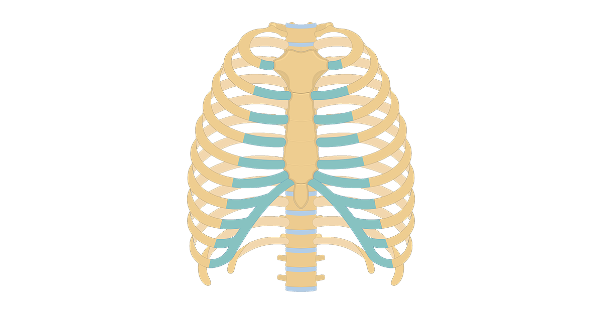
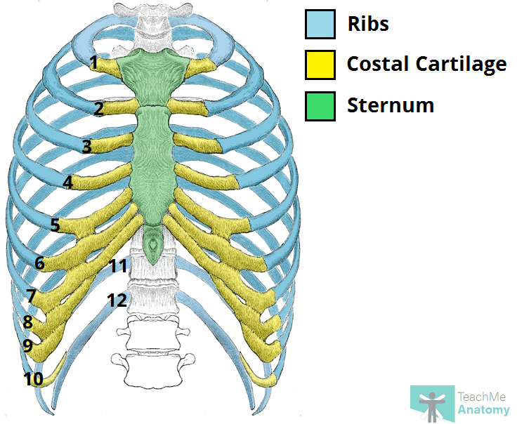
Generally, ribs 1 to 7 are connected to the sternum by their costal cartilages and are called true ribs, whereas ribs 8 to 12 are termed false ribs anatomy of ribs. Cartilage joins adjacent costal cartilage of ribs next to it and the next three ribs.


0 Komentar